PICS~AIC
Authors: Boban P. Abraham, MD, FAAP, FSCAI, FPICS; Athar M. Qureshi, MD, FACC, FSCAI, FAAP, FPICS
How I do it: Tips, Tricks, and Techniques
A PICS Society education series
Percutaneous Axillary Artery Access
The axillary artery access is a convenient alternative access site to femoral artery access in neonates to adults in several situations. The axillary artery is about the same size as the femoral artery and can be used to place large sheaths for interventions. The axillary artery provides more advantageous access and orientation to several anatomic structures and procedures like aortic valve procedures, Blalock Taussig Thomas shunt interventions, patent ductus arteriosus stent interventions and coarctation procedures in addition to mechanical circulatory support. The axillary artery is not an end artery and as such, the risk of limb threatening ischemia is minimal. This is because of the presence of abundant collaterals supplied through the 2nd intercostal artery and subscapular artery. For these reasons, it is beneficial for pediatric and congenital interventional cardiologists to gain experience in obtaining and managing axillary artery access.
This document reviews the anatomy of the axillary artery along with its ultrasonographic and angiographic characteristics. It also describes the techniques to safely access the artery percutaneously and then close the arteriotomy to achieve hemostasis at the end of the procedure.
The axillary artery continues from the subclavian artery and starts at the lateral edge of the first rib, extending to the lateral edge of the Teres Major muscle. The first segment, medial to the pectoralis minor muscle gives rise to the superior (supreme) thoracic artery. The second segment posterior to the pectoralis minor muscle gives rise to the thoracoacromial artery and lateral thoracic artery. The third segment lateral to the pectoralis minor muscle gives rise to the medial and lateral circumflex humeral arteries and subscapular artery. The brachial plexus runs posterior to the second segment of the artery and is an ideal segment for arteriotomy without risk of nerve injury (Figures 1 and 2).
- Identifying the optimal arteriotomy site among the three axillary artery segments
- Avoiding brachial plexus injury
- Preventing pleural entry and pneumothorax
- Managing vessel tortuosity in infants or obese patients
- Achieving hemostasis in a mobile, soft-tissue–rich region
- Preserving critical side branches (e.g., circumflex humeral arteries)
- Accommodating large-bore access for mechanical circulatory support
Access Planning
- Position the patient supine with the arm abducted 90–110° and externally rotated (Figure 3).
- Slight anterior elevation may improve exposure.
- Pre-prep ultrasound helps identify the artery, vein, and branches and optimize arm position.
Imaging – Ultrasound
- Identify pectoralis major/minor, axillary artery and vein, and arterial branches (Figure 4)
- Visualize brachial plexus cords and avoid them (Figure 5)
- Use real-time guidance in both short- and long-axis views.
Imaging – Angiography
- If another access site is available (femoral, radial, brachial), perform an axillary angiogram and leave a catheter or wire in place for guidance (Figure 6, Video 1).
- If not, perform angiography through a micropuncture catheter after initial arteriotomy (Figures 7, 8, Videos 2, 3).
- Confirm the arterial segment and branch anatomy before upsizing.
Optimal Arteriotomy Site
The ideal puncture zone is:
- Between the second and third portions of the axillary artery
- Posterior to or at the lateral border of the pectoralis minor
- Medial to the humeral head
- Across from the coracoid process
- Between:
- Thoracoacromial and lateral thoracic arteries (medially)
- Subscapular and circumflex humeral arteries (laterally)
This location:
- Minimizes brachial plexus injury
- Avoids pleural entry
- Preserves collateral circulation
- Ensures compressibility
- Protects circumflex humeral branches (critical for preventing humeral head avascular necrosis)
Sheaths
- Micropuncture kit
- Appropriately sized sheaths for the planned intervention
Devices
- Perclose sutures for pre-close (large-bore access)
- Covered stents (bailout only)
Other
- High-resolution ultrasound
- Pressure dressing materials
Lateral approach
- Needle puncture in the axilla itself without going through the pectoralis muscles (Figure 9). Used more commonly in infants and younger children and when long-term access is not anticipated.
Medial approach
- Needle puncture in the infraclavicular anterior chest near the deltopectoral groove, through the pectoralis muscles (Figure 10). Used more commonly in older children, adults, obese patient and when long-term access is anticipated, say for mechanical circulatory support.
Real time ultrasonic and fluoroscopic guidance is utilized. Bony landmarks may also be utilized. The optimal axillary arteriotomy site is medial to the humeral head, across from the coracoid process of the scapula (where the pectoralis minor inserts) and lateral to the rib cage.
A micropuncture technique is recommended. An angiogram through the micropuncture catheter is recommended to confirm arteriotomy site prior to upsizing to a sheath.
The optimal arteriotomy site is between the second and third portions of the axillary artery, posterior to or at the lateral border of the pectoralis minor muscle. Once the axillary artery and branches are defined, the access point should be between the thoracoacromial artery and lateral thoracic artery medially and subscapular and the circumflex humeral arteries laterally. This is clinically important because it is associated with the lowest chance of causing brachial plexus injury, does not risk entering the chest and thus reduces the chance of a pneumothorax, and is manually compressible for hemostatic purposes. In the event of needing a covered stent in the axillary artery, this will ensure patency of side branches especially the circumflex humeral arteries whose occlusion can lead to avascular necrosis of the humeral head. Optimal arteriotomy site also preserves collateral supply distally.
Hemostasis
The axillary access site requires meticulous steps to guarantee hemostasis after removal of the sheath. For most infants and pediatric patients and for small bore access, manual compression is sufficient. For large bore access, if there’s sufficient time, two Perclose sutures can deployed at 10 o’clock and 2 o’clock positions in a pre-close fashion. If that is not feasible, a dry closure as described below is recommended. A balloon (size based on the vessel size) is delivered from an alternative access site to the arteriotomy site. Then the sheath is removed and the balloon is gently inflated at the arteriotomy site until oozing stops. Sometimes balloon inflation lasting 15-30 minutes across the arteriotomy site is needed to achieve hemostasis. If the subsequent angiogram shows extravasation at the access site, another prolonged balloon inflation is used. If balloon inflation and manual compression fail, then a self-expanding covered stent can be used as a bail-out strategy.
Applying effective pressure dressing at the axillary arteriotomy site, after manual compression to achieve hemostasis, can be challenging due to lax axillary soft tissue and mobility of the shoulder joint.
A useful technique for axillary artery pressure dressing adapted from 68W Combat Medic Specialist Advanced Individual Training is depicted in Figure 11, Video 4.
The axillary artery is more fragile than the femoral artery making it theoretically more prone to complications. The rate of complications is related to the age and body habitus of the patient, anticoagulation, tortuosity of the artery and size and dwell time of the access. Reported complications include bleeding, hematoma, pseudoaneurysm, dissection, thrombosis, thromboembolism, hand ischemia and compression and injury to the brachial plexus.
The Axillary Access Registry to Monitor Safety (ARMS) Registry (which included 102 consecutive adult patients who underwent transaxillary access for mechanical hemodynamic support across 10 U.S. sites) reported a 15.7% (16/102) rate of procedural complications, 10% of which were minor access site bleeding and hematoma. There were no instances of major bleeding. Other in-hospital complications (occurring >6 hours after the procedure up until discharge) included access site bleeding requiring transfusion (6/102), access site hematoma >4 cm (3/102), neurologic complaints (3/102), pseudoaneurysm formation (1/102), and hand ischemia (1/102) (Reference 1).
In pediatric literature, partial dissection to subclavian artery was noted in 2/20 patients and pseudoaneurysm in 1/20 patients (Reference 5). Pseudoaneurysm formation can be managed by ultrasound guided compression and injection of thrombin. Surgical resection is rarely required in the current era. Arterial dissection (n = 1/70), formation of a hematoma (n = 2/70), and excessive bleeding (n = 2/70) were also noted (Reference 6). Axillary artery occlusive thrombus was noted in 1/6 patients (Reference 7).
Axillary artery access is a versatile and safe alternative to femoral access when performed with careful anatomical understanding, imaging guidance, and meticulous technique. Optimal puncture location—between the second and third arterial segments—minimizes complications and preserves critical collateral circulation. Ultrasound and angiography are essential for safe access, and thoughtful hemostasis strategies ensure reliable closure even after large-bore interventions.
- Seto A, Estep J, Tayal R, et al. SCAI Position Statement on Best Practices for Percutaneous Axillary Arterial Access and Training. Journal of the Society for Cardiovascular Angiography & Interventions, 2022; 1.
- Amir Kaki, M. Chadi Alraies, Theodore L. Schreiber. Peripheral Matters – Axillary Artery: Alternate Access for Large Bore Interventional Procedures. Cardiology Interventions Jun 22, 2018.
- Davenport JJ, Lam L, Whalen-Glass R, Nykanen DG, Burke RP, Hannan R, et al. The successful use of alternative routes of vascular access for performing pediatric interventional cardiac catheterization. Catheter Cardiovasc Interv. (2008) 72(3):392–8. 10.1002/ccd.21621
- Martin RP, Qureshi SA, Arnold R. Percutaneous balloon aortoplasty of recoarctation: an alternative approach using the axillary artery. Int J Cardiol. (1989) 22(1):119–21. 10.1016/0167-5273(89)90144-7
- Breatnach CR, et al. Percutaneous axillary artery approach for ductal stenting in critical right ventricular outflow tract lesions in the neonatal period. Catheter Cardiovasc Interv. 2019 Jun 1;93(7):1329-1335.
- Schranz D, et al. Axillary artery access for cardiac interventions in newborns. Ann Pediatr Cardiol. 2008 Jul-Dec; 1(2): 126–130.
- Bauser-Heaton H, Qureshi AM, Goldstein BH, Glatz AC, Nicholson GT, Meadows JJ, et al. Use of carotid and axillary artery approach for stenting the patent ductus arteriosus in infants with ductal-dependent pulmonary blood flow: a multicenter study from the congenital catheterization research collaborative. Catheter Cardiovasc Interv. (2020) 95(4):726–33
Figure 1
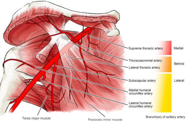
Figure 1 Legend: Branches of the axillary artery and their relationship to the pectoralis minor and teres major muscles, grouped by segment (medial, behind, lateral).
Figure 2
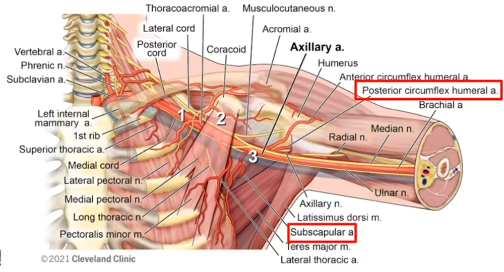
Figure 2 Legend: The three segments of the axillary artery (1, 2, 3) with the surrounding brachial plexus cords and branch vessels. The subscapular and posterior circumflex humeral arteries are highlighted.
Figure 3
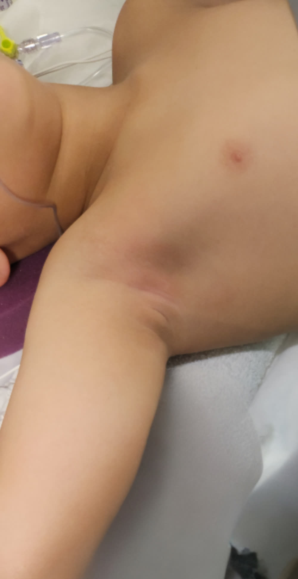
Figure 3 Legend: Patient positioning — supine with the arm abducted 90–110° and externally rotated.
Figure 4
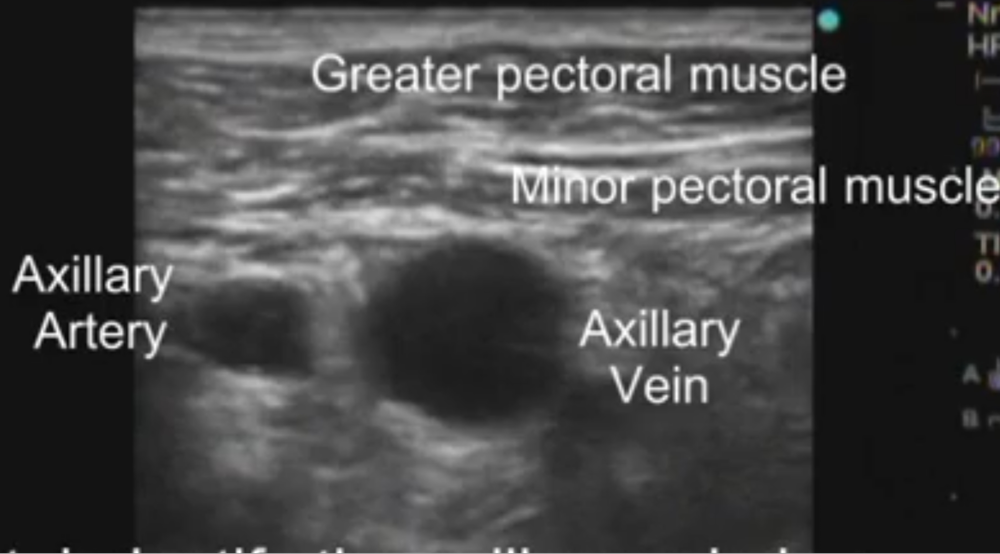
Figure 4 Legend: Ultrasonographic identification of the pectoralis major and minor, the axillary artery and vein, and the arterial branches.
Figure 5
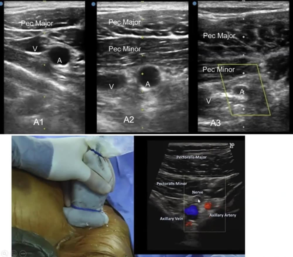
Figure 5 Legend: A1, A2 and A3 represent ultrasonographic appearance of the first, second and third segments of the axillary, respectively. In A3, the probe is placed lateral to the deltopectoral groove and is too lateral for optimal arteriotomy. Note the cord of the brachial plexus, as noted by the designation, Nerve, anterior to the artery and vein. Courtesy of SCAI position statement, 2022 (Reference 1).
Figure 6
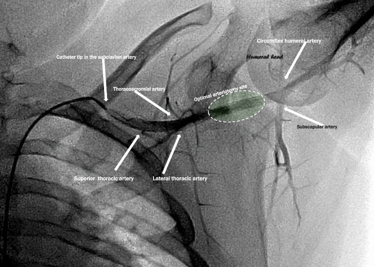
Figure 6, Video 1: A catheter introduced from the femoral artery is utilized to perform axillary artery angiogram.
Figure 7
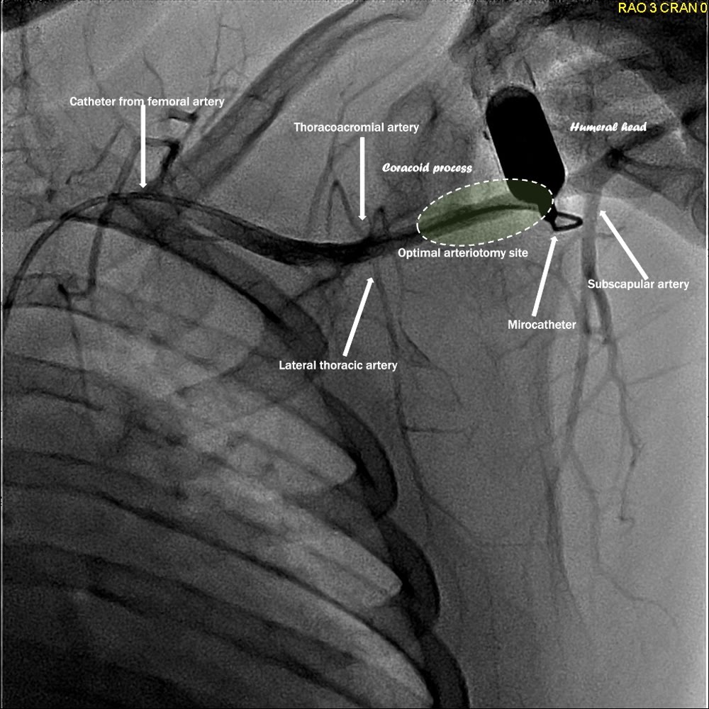
Figure 7, Video 2: The catheter introduced from the femoral artery is in the axillary artery. The angiogram was performed utilizing a micropuncture catheter in the axillary arteriotomy site. The arteriotomy site is optimal between the thoracoacromial artery and lateral thoracic artery proximally and subscapular artery distally, across from the coracoid process and medial to the humeral head.
Figure 8
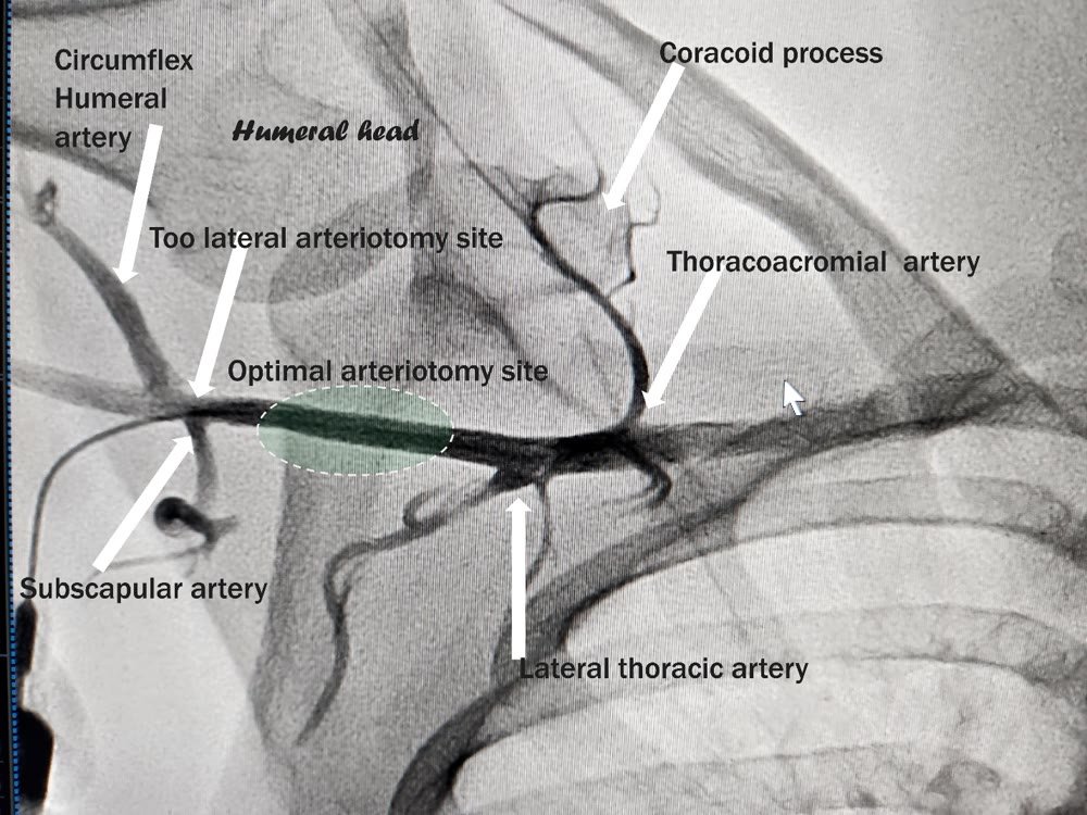
Figure 8, Video 3: The angiogram was performed utilizing a micropuncture catheter in the axillary arteriotomy site. The arteriotomy site is suboptimal, too distal and in proximity to the subscapular and circumflex humeral arteries.
Figure 9
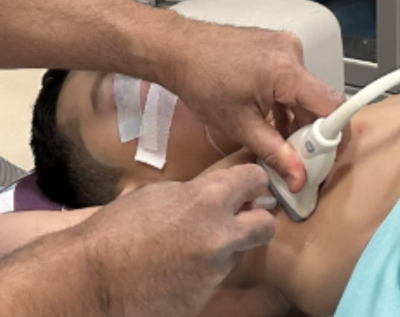
Figure 9 Legend: Lateral approach — needle puncture in the axilla itself, without traversing the pectoralis muscles, under real-time ultrasound guidance.
Figure 10
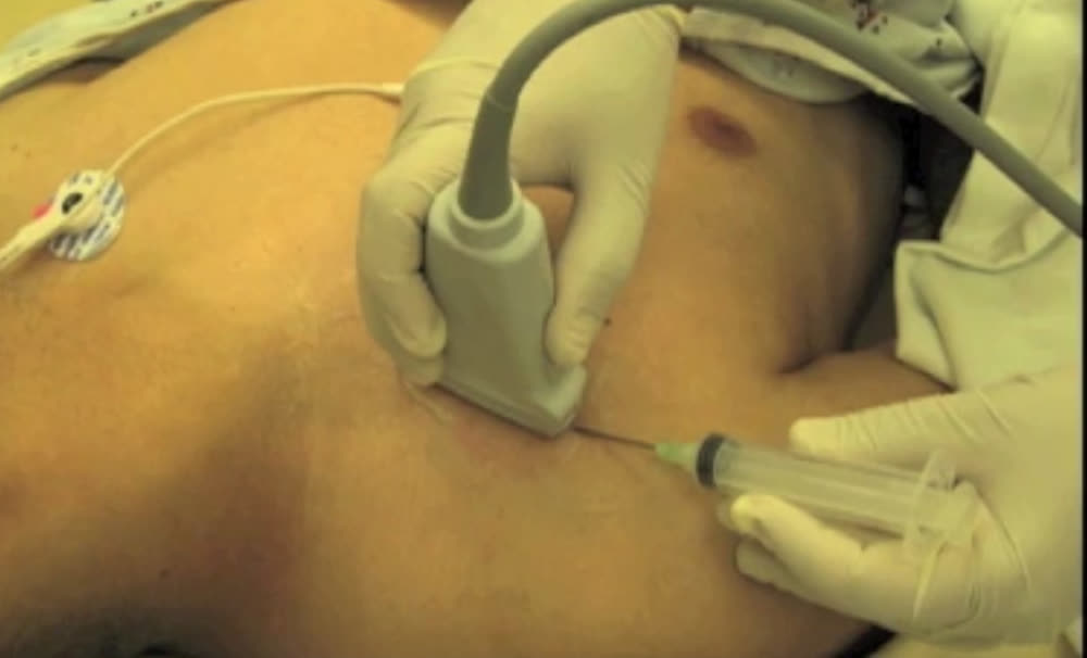
Figure 10 Legend: Medial approach — needle puncture in the infraclavicular anterior chest near the deltopectoral groove, through the pectoralis muscles, under real-time ultrasound guidance.